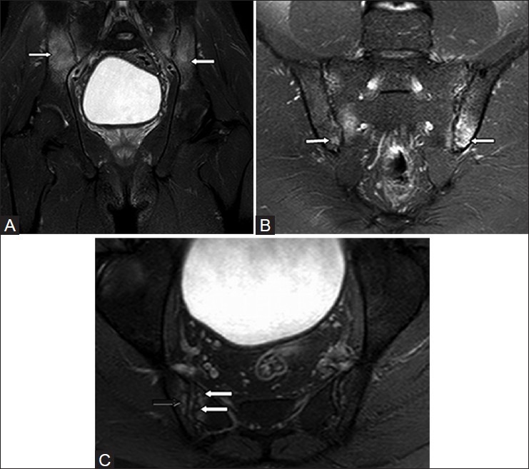
Figure 6 (A-C).

Findings of acute sacroiliitis on MRI – subchondral edema and erosions. (A) Coronal STIR MR image in a 21 year old female with ankylosing spondylitis shows bilateral symmetrical edema in the subchondral and inferior aspects of the sacroiliac joints (white arrows). (B) Oblique coronal STIR MR image of the sacroiliac joint shows bilateral symmetrical edema in the posteroinferior aspect of the joints (white arrows). (C) Axial STIR MR image through sacroiliac joints shows multiple erosions (white arrows) with subchondral edema (black arrow)
