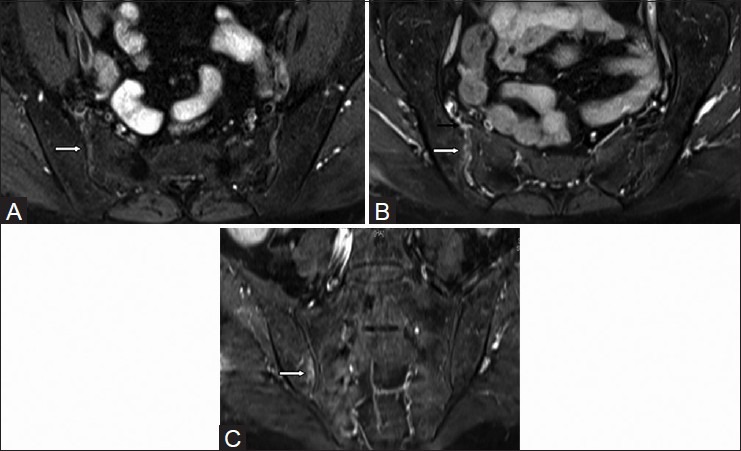
Figure 8 (A-C).

Findings of acute sacroiliitis on MRI - synovitis and marrow enhancement. Axial T1W fat-saturated (A) pre-contrast and (B) post-contrast MR images in a 52 year old female with psoriasis show enhancing joint synovium on the right side consistent with synovitis (white arrows); enhancement of anterior capsule is also seen (black arrow). (C) Coronal T1W fat-saturated post-contrast MR image shows subchondral bone marrow enhancement in the right iliac bone, findings consistent with active osteitis (white arrow)
