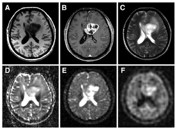
Fig. 11.
Proton and sodium images from a patient with glioblastoma (WHO grade IV) of the left medial frontal lobe. (A) T1 weighted MRI, (B) T1 weighted MRI with contrast medium (rim enhancement), (C) T2 weighted MRI showing cystic and solid portions of the lesion and perifocal brain edema, (D) DWI showing elevated ADC values in the center of the tumor, (E) sodium MRI showing increased sodium signal in the tumor, (F) sodium MRI with fluid suppression by inversion recovery (IR), also showing increased sodium signal mainly at the center of the tumor. Proton images (A–D) were acquired at 3 T while sodium images (E and F) were acquired at 7 T. Reproduced with permission from Ref. [135]. Coyright 2011, Wolters Kluwer Health.

