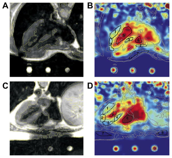
Fig. 14.
Comparison between 1H and 23Na cardiac images at 1.5 T of healthy volunteers. Registered axial 1H MRI FSE images (A and C), cardiac-gated at 60–70 bpm, echo train length = 8, effective TE = 17 ms. Coil sensitivity adjusted axial slices from three-dimensional 23Na TPI data sets of the heart (B and D) with TR/ TE = 100/0.17 ms, and adiabatic half passage (AHP) excitation from healthy human volunteers. The 23Na image intensity was adjusted for the coil receiving sensitivity using a computer-generated B1 field estimate of the coil. The 1H image data were interpolated in three dimensions so that images (A and C) match the spatial positions of the images (B and D). The resulting in-plane resolution was 128 × 128, at the same slice spacing as the 23Na data. The three-dimensional 23Na image data were interpolated to 128 × 128 pixels in-plane to facilitate copying of contours and ROI. Yellow contours are drawn in the 1H images (A and C) and copied to the 23Na images (B and D) (shown as gray lines) to guide placement of the ROIs (black lines in the 23Na images). ROIs were placed in the lateral LV wall, anterior LV wall, and septum (ROI numbers 1–3 or 1–4 in all images). Additional ROIs were placed in the left ventricle for LV blood (ROI number 5) and in the right ventricle for RV blood (ROI number 6). Reproduced with permission from Ref. [174].

