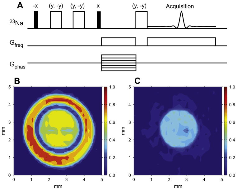
Fig. 5.
2D 23Na images obtained with the pulse sequences shown in (A) – the quadrupolar jump-and-return (QJR) sequence. (B) The signal is excited by a hard 90° pulse. (C) The QJR sequence with a delay of 2.5 ms was used to excite the signal. The phantom consists of two concentric tubes (3 mm and 5 mm od, respectively), the inner tube filled with Pf1 bacteriophage solution (fQ = 205 Hz) and an outer with 50 mM NaCl solution. Reproduced with permission from Ref. [109].

