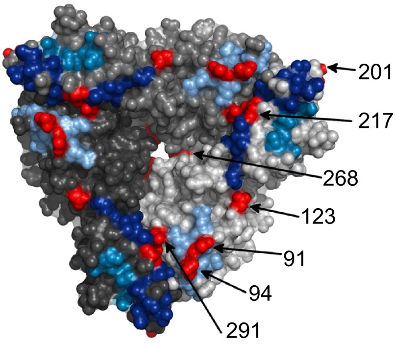
Figure 1.
A surface representation of the VP7 trimer (PDB ID: 3FMG). Each trimer is colored a different shade of gray and the antigenic regions A, C and F are colored light blue, dark blue and mid blue, respectively. The surface exposed amino acid residues that differ between the 2010 G1P[8] Alice Springs outbreak strain and Rotarix® vaccine strain are shown in red.

