Figure 3.
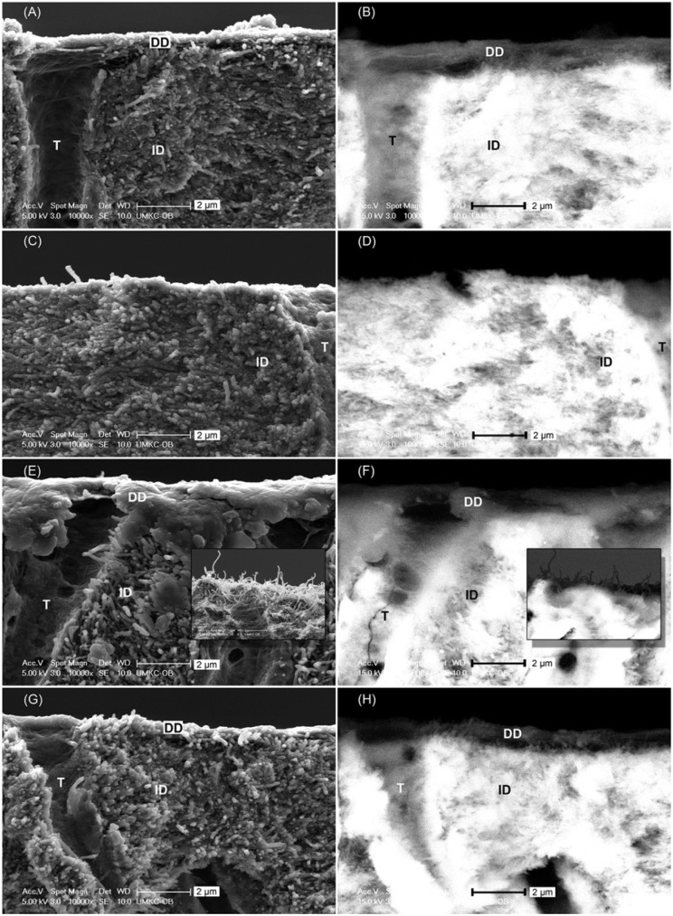
Scanning electronic microscopy images of cross section of fractured dentin slabs under (A, C, E, G) secondary electron mode and (B, D, F, H) backscattered electron mode. (A, B) Typical morphology of etched dentin predigestion, regardless of cross-linkers. (C, D) Glutaraldehyde–phosphoric acid–etched dentin after 1 hr of digestion, showing no demineralized layer left. (E, F) GSE20-etched dentin after 1 hr of digestion, featuring a thin demineralized film draping over the edge and hair-like morphology on the complementary piece (insets). (G, H) GSE10- and GSE5-etched dentin after 1 hr of digestion, showing a morphology unchanged from predigestion. DD, demineralized dentin; GSE, grape seed extract; ID, intact dentin; T, tubule.

