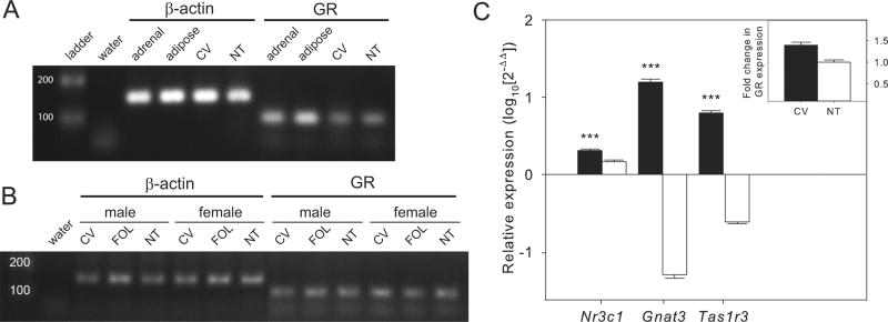
Fig. 1.

Expression of glucocorticoid receptor (GR) mRNAs in taste and other tissues from mice using RT-PCR. (A) Both circumvallate (CV) taste papillae and non-taste (NT) tissue express GR mRNAs. The water lane is a negative control, adrenal and adipose tissues from mice were used as positive tissue controls for GR expression and β-actin is a positive control gene. (B) Expression of GR was seen in all taste (CV, foliate papillae [FOL]) and NT tissues in both sexes. (C) Gene expression of GR (Nr3c1) is enriched in CV compared to NT based on quantitative PCR (n = 13 mice). Gnat3 and Tas1r3 are positive control genes for taste tissues. Gene expression was relative to control gene expression (β-actin) within each sample. Inset shows fold change in GR expression for CV compared to NT. Bars represent average ± SEM, asterisks indicate statistically significant differences (P < 0.001).
