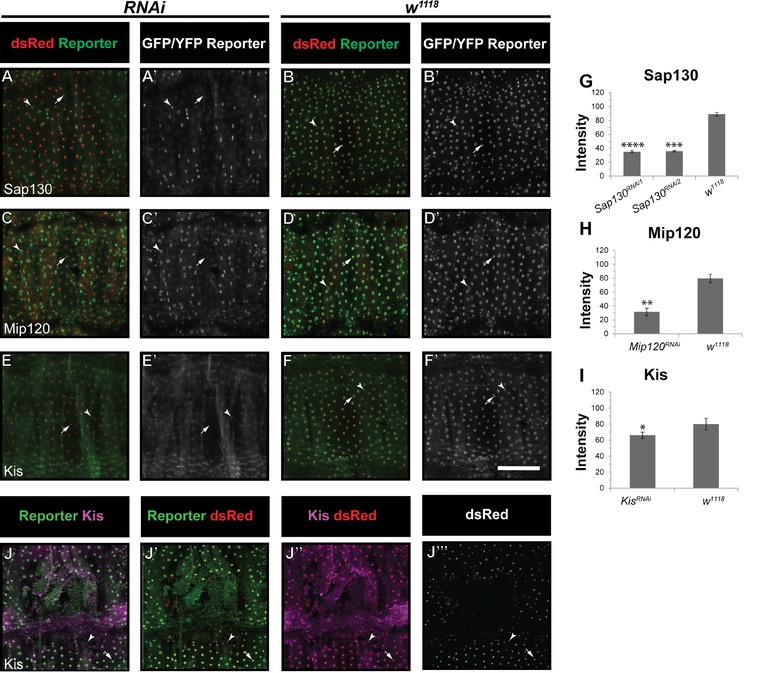
Figure 2.

RNAi knockdown reduces expression of the Sap130, Mip120, and Kis reporters. (A)−(F′) Unwounded segments of dissected epidermal whole mounts of larvae heterozygous for an epidermal Gal4 driver (see Table 1), UAS‐DsRed2nuc, the indicated reporter, and the indicated UAS‐RNAi. Larvae were raised at 30°C. (A)−(A′) Sap130 reporter with UAS‐Sap130 RNAi1. (B)−(B′) Sap130 reporter with w 1118. (C)−(C′) Mip120 reporter with UAS‐Mip120 RNAi. (D)−(D′) Mip120 reporter with w 1118. (E)−(E′) Kis reporter with UAS‐Kis RNAi. (F)−(F′) Kis reporter with w 1118. Arrowheads, muscle nuclei that do not express dsRed2Nuc; arrows, epidermal nuclei; note the lack of GFP staining in (A)−(A′), (C)−(C′), and (E)−(E′) compared with controls. (G), (I) Quantification of reporter intensity in epidermal nuclei of indicated genotypes. *P < 0.05; **P < 0.005; ***P < 0.001; ****P < 0.0005 (Student's t test). For the remaining reporter/RNAi combinations tested no statistically significant reduction in signal was observed. (J)−(J′′′′) Kis antibody staining (J, J′′, J′′′′) is reduced at the wound edge 4 h post‐wounding (J′′), and overlaps with Kis reporter staining (J). Scale bar 200 μm.
