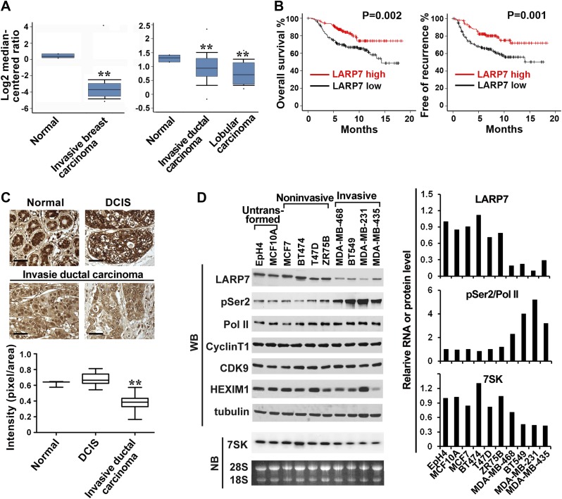
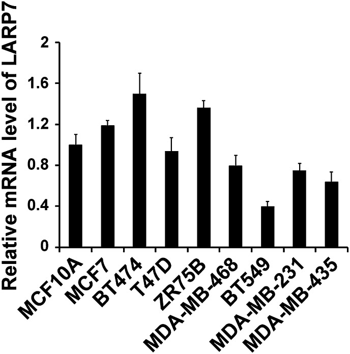
Figure 1. LARP7 is significantly downregulated in invasive human breast cancer tissues and cells.
(A) Box plots show decreased levels of LARP7 in invasive breast carcinoma (left), invasive ductal carcinoma, and lobular carcinoma (right) compared with normal breast tissues in two microarray data sets. **: the p values (p<0.01, compared with normal breast tissues) were determined by the Student's t test. (B) Kaplan–Meier analysis of overall survival and recurrence-free survival of breast cancer patients stratified by the expression of LARP7. The p values were calculated by the log-rank test. (C) Immunohistochemical staining of LARP7 in normal human mammary tissue (n = 6), ductal carcinoma in situ (DCIS) (n = 14), and invasive ductal carcinoma (n = 120). The intensity of LARP7 staining was quantified using ImageJ Plus and shown in the box plot below. Scale bars represent 40 μm. **: the p value (p<0.01, compared with normal breast and DCIS tissues) was determined by the Student's t test. (D) Western blotting (WB) analysis of the levels of LARP7, phospho-Ser2 (pSer2), total Pol II, CyclinT1, CDK9, and HEXIM1 in various breast cancer cell lines (upper panels) and Northern blotting (NB) analysis of 7SK snRNA levels (lower panels). Tubulin, 28S and 18S RNAs were used as loading controls. Expression of LARP7, pSer2 of Pol II, and 7SK RNA was quantified, normalized to that in EpH4 cells, and shown in the graph to the right.


