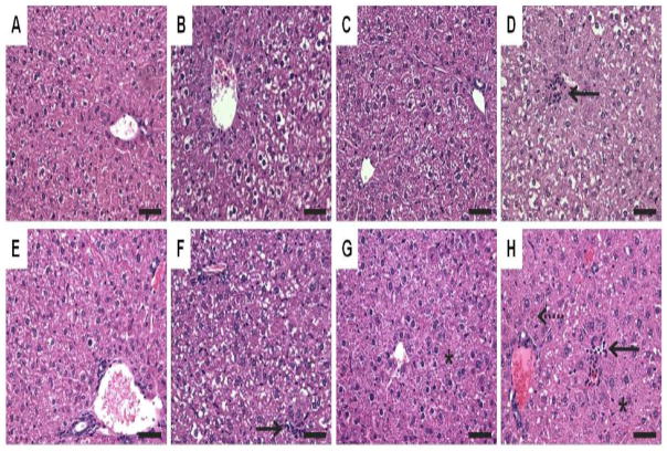
Figure 3.
Standard hematoxylin and eosin staining of liver sections at 24 (top panel: A–D) and 168 h (bottom panel: E–H). (A&E) Sesame oil elicited minimal vacuolization. (B&F) 30 μg/kg TCDD elicited vacuolization and inflammation (solid arrow). (C&G) 300 mg/kg PCB153 resulted in minimal vacuolization and hypertrophy (asterisk). (D&H) MIX resulted in vacuolization, inflammation (solid arrow), hypertrophy (asterisk) and necrosis (dashed arrow). Bars = 50 μm.

