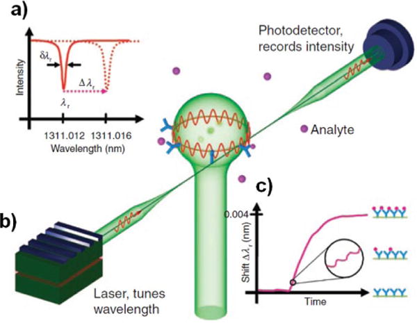
Figure 4.

Illustration of a whispering gallery mode biosensor. Light is coupled into a microsphere evanescently via a tapered optical fiber. Certain wavelengths of light will resonate in the cavity, causing dips in transmission through the optical fiber at those wavelengths. Binding of molecules on the cavity surface will cause a shift in the resonant frequency spectrum of the cavity, and thus the resonant frequency dips monitored at the detector will shift. As more analyte binds to the surface, this resonant frequency shift will increase.81 Reprinted by permission from Macmillan Publishers Ltd: Nature Methods. Vollmer F, Arnold S. Whispering-gallery-mode biosensing: label-free detection down to single molecules. Nat Meth 2008, 5:591–596. Copyright 2008.
