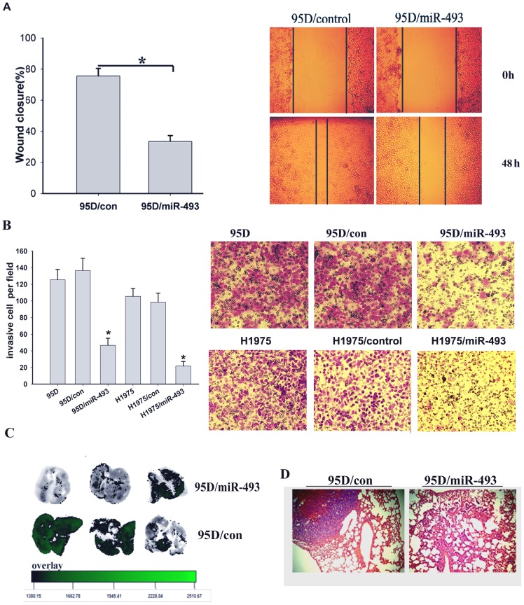
Figure 3. Expression of miR-493 inhibits the migration and invasion of lung cancer cells.
A, scratch wound assays were conducted on 95D cells transfected with miR-493 and its paired control. The results from 3 separate assays were averaged together and graphed. *, P<0.05 (left). Representative images of the assays are shown. Original magnification: ×200 (right). B, the invasive properties of the H1975 and 95D cells were analyzed with a Transwell assay using a Matrigel-coated chamber. Migrated cells were plotted as the average number of cells per field of view from 3 different experiments, as described in the Materials and Methods. *P<0.01 (left). Representative images of the assays are shown. Original magnification: ×200 (right). C, in vivo metastasis assays were used to examine the lung metastatic ability of 95D/miR-493 cells labeled with green fluorescent protein(GFP) and its paired control cell 95D/control. Lung cancer cells were injected into the tail veins of five week- old mice. On day-28, all animals were sacrificed and the lungs were excised. The lung metastasis images were obtained with a Bruker Small Animal Imaging System. The lung tissue was then removed, fixed, paraffin-embedded, serially sectioned, and subjected to hematoxylin and eosin (H&E) staining. Left, Representation of the detected GFP signal in each of the five animals (the images were overlaid with green fluorescence and white light). Right, The metastatic field and fluorescence intensity significantly differed between the 95D/miR-493 group and the control group. D, Histological examination of pulmonary metastases from 95D/miR-493 and 95D/control cells by haematoxylin and eosin staining.

