Figure 4. Cell distribution at the initial seeding.

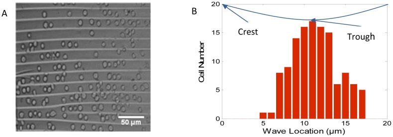
(A) Microscope image of cells on a wavy surface with 20 µm spacing and 6.6 µm height; (B) The number of endothelial cells at different wave locations.

(A) Microscope image of cells on a wavy surface with 20 µm spacing and 6.6 µm height; (B) The number of endothelial cells at different wave locations.