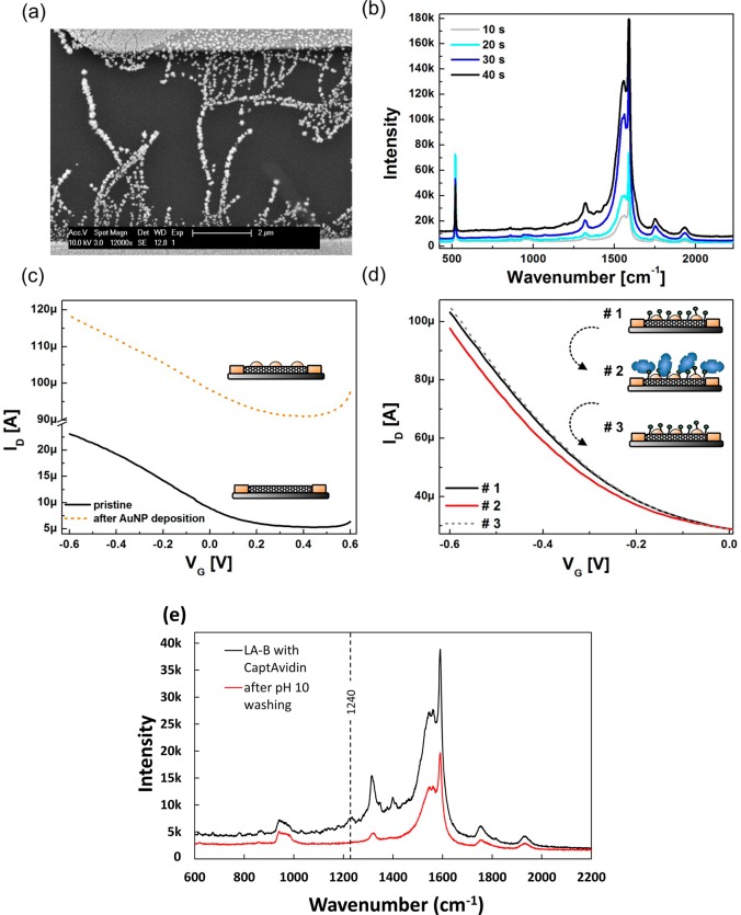
Figure 3.
(a) SEM image of gold nanoparticle decorated SWNTs. The particle sizes obtained from the applied parameters for this device are 50 to 200 nm. (b) Raman spectra of gold nanoparticle decorated SWNTs using varying deposition times of bulk electrolysis (deposition voltage −0.4 V). (c) Transfer characteristics of a SWNT FET recorded before and after functionalization with gold nanoparticles. (d) Transfer characteristics recorded for CaptAvidin detection with a LA-B functionalized device. (1) Transfer curve was taken before exposure to CaptAvidin, (2) after incubation with 140 nM CaptAvidin, and (3) after 15 min exposure to pH 10 buffer. (e) Raman spectra of gold nanoparticle-decorated SWNTs functionalized with LA-B after incubation with CaptAvidin and after pH 10 washing. Peaks unique to the protein can be discerned after incubation which disappear upon rinsing.

