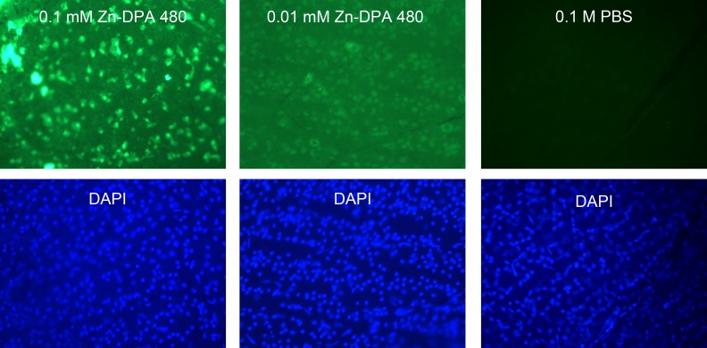
Figure 2.
Concentration effect of Zn-DPA 480 probes at 4 hours after injection of NMDA. The solutions of Zn-DPA 480 were injected 1 hour before euthanasia. Cells in the RGC layer were labeled by both 0.1 and 0.01 mM Zn-DPA 480 concentrations. Intravitreal injection of 0.1 mM Zn-DPA 480 labels the RGC layer cells with a stronger signal than 0.01 mM after NMDA injection; 1 mM Zn-DPA 480 gave similar signal intensity as 0.1 mM Zn-DPA 480 (data not shown). There was no labeling in PBS-vehicle, instead of NMDA, injected eye. To compare labeling intensity, all photographs were taken under the same magnification at a fixed 10-second exposure time.

