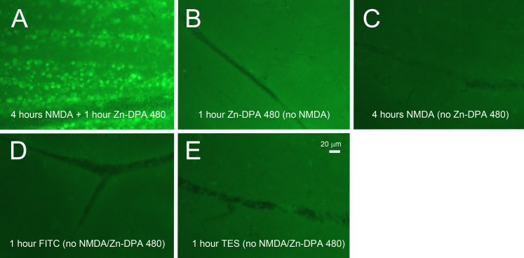
Figure 3.
Fluorescence intensity on retinal wholemount at 1 hour postinjection of Zn-DPA 480 imaging probes. (A) At 4 hours after 40 mM NMDA injection, positive cells were labeled by 0.1 mM Zn-DPA 480 and background fluorescence was increased. There were no positive cells in the retinas after injection of (B) Zn-DPA 480, (C) NMDA, (D) FITC, and (E) TES vehicles, and the background intensities were noticeably lower than the retina treated with Zn-DPA 480 and NMDA (A). The exposure time was 10 seconds.

