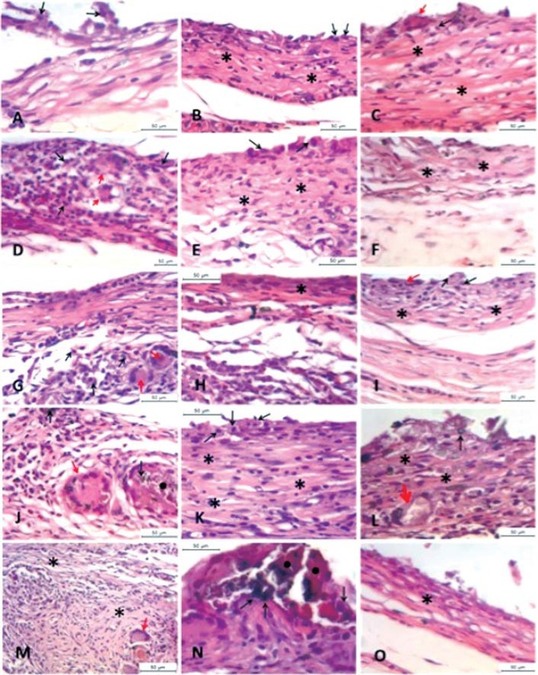
Figure 1.
(A-C) Group 1 (0%WPC); (D-F) Group 2 (15%WPC); (G-I) Group 3 (20%WPC); (J-L) Group 4 (30%WPC); (M-O) Group 5 (50%WPC). At 15 days, a chronic inflammatory infiltrate, moderate with predominance of macrophage (black arrow) is observed, presence of giant multinuclear cell (red arrow), areas with Portland cement particles involved with giant cells (black circle) and a mild presence of fiber connective tissue (asterisk) in all groups. At 30 days, a decrease in the inflammatory infiltrate and a discrete increase in fiber connective tissue (asterisk) in comparison with the 15 days period were observed. Futhermore, at 30 days the presence of macrophage cells (black arrow) is predominant. Areas with Portland cement particles involved with giant cells are still present in this period (black circle). At 60 days, the chronic inflammatory response is mild with isolated macrophage-like cell (black arrow) and giant cells (red arrow). The presence of fiber connective tissue related to the repair process is evident (asterisk)

