Figure 2.
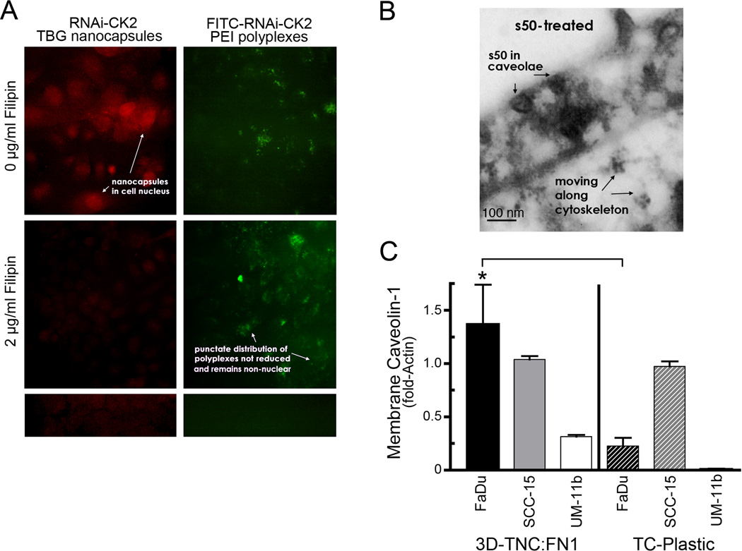
Uptake of the s50-TBG nanocapsule is mediated via caveolae/lipid raft pathway. A, SCC-15 cells were first treated with 2 µg/ml filipin to disrupt caveolae (middle panels), then with 1 µg/ml s50-TBG-RNAi-CK2 (left panels) or 5 µg/ml FITC-labeled RNAi-CK2-PEI polyplexes (right, top and middle). Nanocapsules (red) were indirectly detected by fluorescent anti-sheep IgG (left, top and middle). RNAi-CK2-PEI polyplexes were directly visualized via the FITC-label (right). No-primary antibody control is shown at bottom left and untreated control at bottom right. Original magnification, 40,000×. B, transmission electron micrograph of nanocapsules in surface caveolae of SCC-15 cells grown on TNC:FN1 and treated with 20 µM s50-TBG-siCK2. Original magnification, 45,000×. C, membrane-associated caveolin-1 expression is upregulated in HNSCC lines grown on TNC:FN1 coated nanofibers versus standard tissue culture (TC) plasticware. Mean ± SE are shown. *p<0.0001.

