
Figure 1.
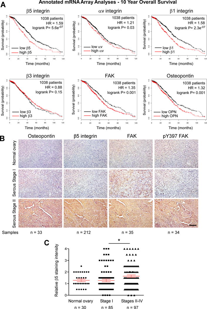
Association between OPN, β5 integrin, and FAK activation in serous ovarian cancer. A, Kaplan-Meier analyses of integrin β5, αv, β1, β3, FAK, and OPN mRNA levels in 1038 patient samples. High (red) versus low (black) mRNA expression shows patient overall survival probability over 120 months. Hazard ratio (HR) and logrank P significance values are shown (inset). B, representative immunohistochemical staining of sections obtained from paraffin-embedded normal ovary, serous Stage I, and serous Stage II ovarian tumor tissue arrays using antibodies to pY397 FAK, total FAK, β5 integrin, and OPN. Scale is 100 μm. C, β5 integrin staining intensity (0–4) in annotated ovarian tissue arrays. Values are means (+/− SEM, * p < 0.05, n= sample number).