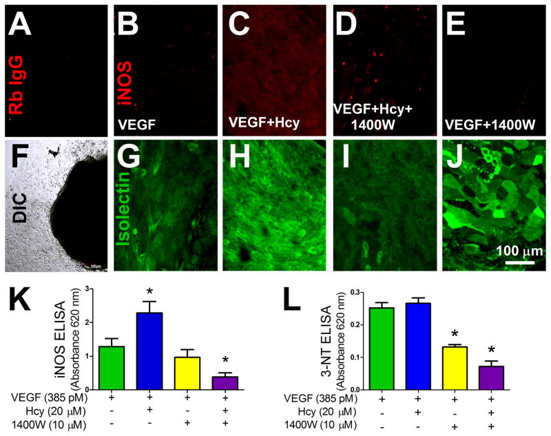
Figure 5. iNOS Expression in Endothelium with Hcy Treatment.
(A) Outgrowth after 4 days of treatment (VEGF 385 pM) labeled with a non-specific rabbit IgG antibody and anti-rabbit Alexa Fluor 647 (red). Fluorescence image (A) of the same area of growth shown in the DIC image (F). (B–E) iNOS rabbit IgG antibody labeling (red) with corresponding isolectin labeling (green) (G–J) of the same area of growth. For imaging of both the isolectin and the iNOS labeling, laser power, offset, and intensity levels were held constant across conditions. Outgrowth after 4 days of treatment (VEGF 385 pM, Hcy 20 μM, and 1400W 10 μM). (K) iNOS expression in a monolayer of microvascular endothelial cells after indicated treatment, VEGF group n=28, VEGF+Hcy group n=18, VEGF+1400W group n=13, VEGF+Hcy+1400W group n=29, * p < 0.05 from VEGF. (L) Quantification of 3-NT after treatment with 1400W, VEGF group n=4, VEGF+Hcy group n=5, VEGF+1400W group n=5, VEGF+Hcy+1400W group n=12, * p < 0.05 from VEGF group.

