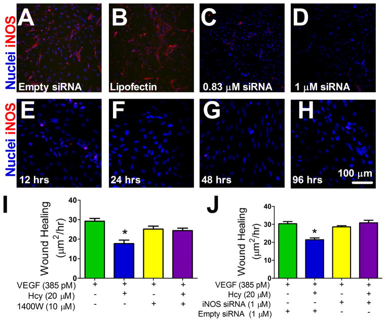
Figure 7. Effect of Hcy and iNOS Activity on Monolayer Endothelial Outgrowth.
(A–D) siRNA dose response, Hoechst nuclear stain (blue) and iNOS labeling (red). Panel A is a representative image of a monolayer of cells with the empty siRNA vector and panel B is a representative image of the transfection agent, lipofectin, alone. (E–H) siRNA time response after an initial 24 hr transfection period. A final dose of 1μM of siRNA was used for wound healing experiments which did not exceed 96 hrs from transfection. (I–J) Confluent microvascular endothelial cells were scrape-wounded and the initial wound area was measured. Wound area was measured again at 48 hrs. The difference between initial wound area and final wound area was divided by time to quantify the rate of outgrowth into the denuded area between groups. (I) Rate of outgrowth into the denuded area with VEGF 385 pM, Hcy 20 μM, and 1400W 10 μM as treatments as indicated, n=12, * p < 0.05 vs. VEGF group. (J) Wound healing rate with VEGF 385 pM, Hcy 20 μM, and 1 μM siRNA or an empty siRNA vector as indicated. VEGF+Empty siRNA group n=6, VEGF+Hcy+Empty siRNA group n=5, VEGF+siRNA group n=5, VEGF+Hcy+siRNA group n=6, * p < 0.05 vs. VEGF+Empty siRNA group.

