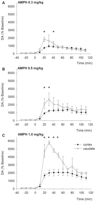
Figure 2.
The increase in extracellular dopamine levels exhibits a different temporal profile across regions. Upon IV injection of AMPH, DA levels in the cortex (filled symbols) increased slowly, which was followed by a slow decline. In the caudate (open symbols), levels increased rapidly, followed by an initial rapid, and subsequent slower, decline. *p<0.05 between cortex and caudate

