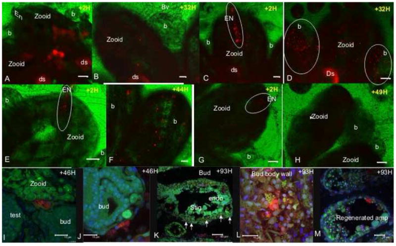
Fig 2. In vivo labeling and tracing of cells distribution in Botryllus colonies.

A. A zooid with hundreds of in situ labeled cells (red) in its body wall, 2 hours post labeling. B. Same zooid, 32 hours post labeling, labeled cells are observed in the zooid but not in its buds. C. Labeled cells in the zooid body wall and the EN (outlined), 2 hours post labeling. D. Homing of labeled cells from the endostyle in panel C to remote buds in the colony, 32 hours following labeling. Labeled cells are detected in the buds (outlined). E. A few labeled cells in the zooid EN (outlined), 2 hours post labeling. F. Labeled cells in the buds 44 hours post labeling. These buds were directly connected to the EN labeled zooid shown in panel E. G. Labeled cells which were transplanted from the vasculature into the zooid EN (outlined), 2 hours post labeling. H. Same zooid 49 hours post labeling, labeled cells taken from vasculature, did not increase in number and did not home to developing buds. I. and J. A few labeled cells in 2 different buds (red), 46 hours post EN labeling (confocal images). K. 93 hours following EN labeling, labeled cells are detected in the bud’s body wall and stigmata (arrows; confocal image). L. Higher magnification confocal image of labeled cells in the bud’s body wall from panel K. M. Labeled cells in the regenerating vasculature epithelial 93 hours following EN labeling and vasculature removal (confocal image). Vybrant Did labeling -red, Hoechst nucleus stain-blue, natural autofluorescence (501nm emissions spectra) –green. EN-endostyle niche, endo-endostyle, b-bud, ds-digestive system, st-stigmata, amp-ampulla, bv-blood vessel, H-hour. scale bar A-H=100μm, I, J, L =15 μm, K-50 μm, M=25μm.
