Fig. 3.

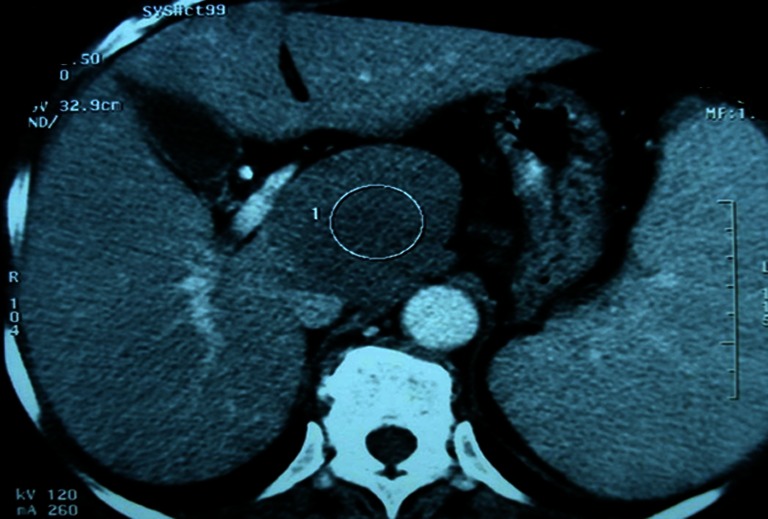
Post-contrast abdominal CT scan shows the lesion with a well-demarcated margin taking just the contrast with peripheral vascular enhancement

Post-contrast abdominal CT scan shows the lesion with a well-demarcated margin taking just the contrast with peripheral vascular enhancement