Figure 2.
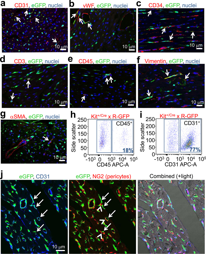
Analysis of cardiac cells from Kit+/Cre × R-GFP mice. a, b, c, d, e, f, g, Immunofluorescent images of heart histological sections from Kit+/Cre × R-GFP mice at 4 weeks of age stained with eGFP antibody (green), nuclei in blue and either CD31, von Willebrand factor (vWF), CD34, CD3, CD45, vimentin or smooth muscle α-actin (αSMA) in red. Arrows show cells with overlap in staining. h, i, FACS plot showing lineage markers of heart isolated c-kit derived eGFP+ cells for CD45 (h) and CD31 (i) (representative of n=6 for CD45 at 4 weeks of age, and n=3 for CD31 at 12 weeks of age). j, Immunofluorescent image from heart histological section of a Kit+/Cre × R-GFP mouse at 4 weeks for eGFP fluorescence (green), CD31 antibody staining (blue) and NG2 antibody staining (red). Right panel shows composite with transmitted light.

