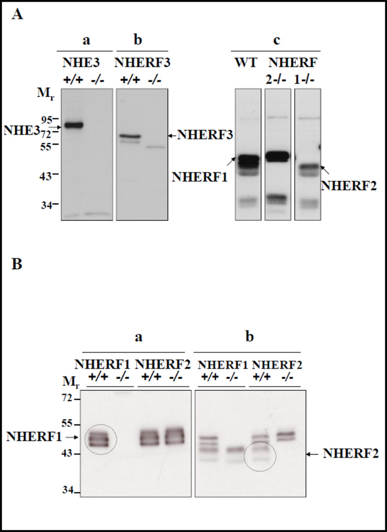
Fig. 1.
Expression of NHE3 and NHERFs in murine BBM. A) Western blots depicting antibody specificities of a) Antibody against NHE3 (~87 KD) in BBM of Nhe3+/+ and Nhe3−/− mice, b) antibody against NHERF3 (~72 KD) in BBM of Nherf3+/+ and Nherf3−/− mice, and c) the rabbit antibody against NHERF2 cross reacts with NHERF1 as depicted by double bands seen at ~48 KD for NHERF1 and ~46 KD for NHERF2 in the WT BBM. The ~46 KD bands were absent in Nherf2−/− BBM, and the ~48 KD bands were absent in Nherf1−/− BBM. B) This blot displays a different BBM preparation from NHERF1 KO and respective WT, as well as NHERF2 KO and the respective WT, probed with the anti- NHERF1 antibody (left panel) and the anti-NHERF2 antibody (right panel). Exposure times were 3 seconds (left panel) and 1 minute (right panel). See explanation in text. It is evident that the anti-NHERF1 antibody stains three bands, all of which are absent in NHERF1 KO BBM, and does not cross react with NHERF2. The NHERF2 antibody crossreacts with the lower two bands stained by anti-NHERF1.

