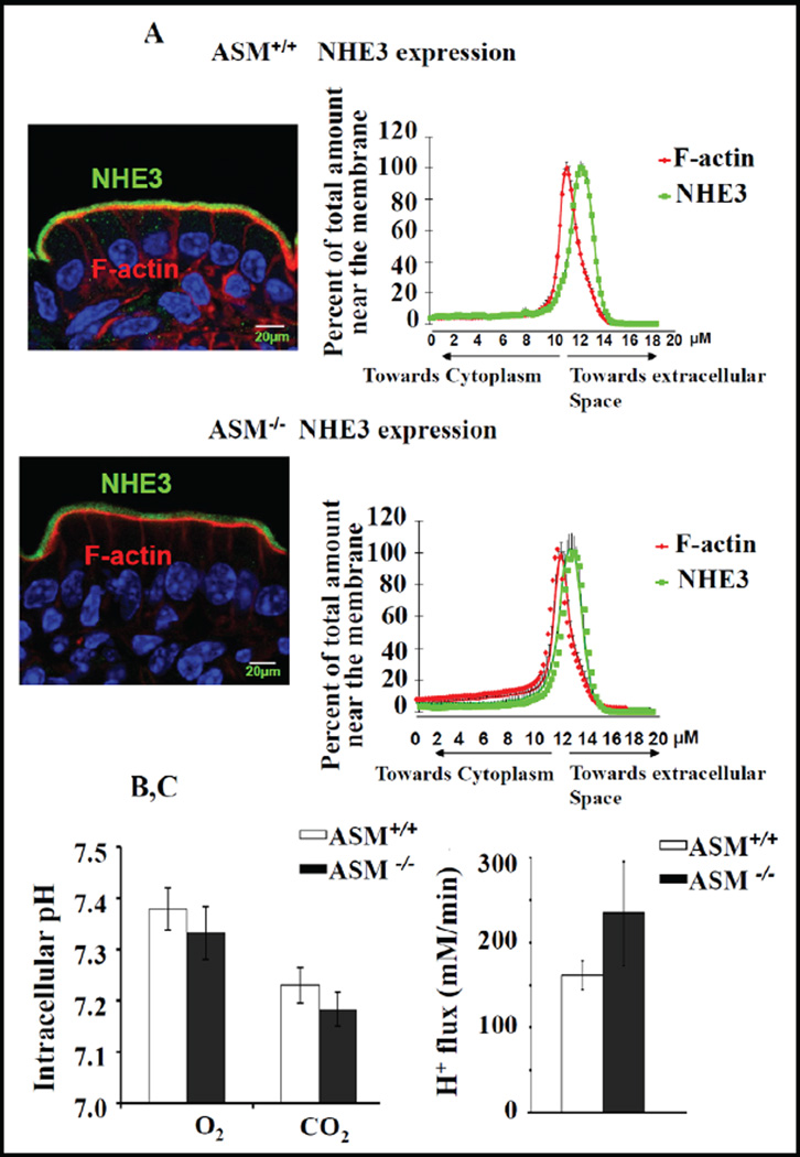
Fig. 8.
NHE3 BBM localization and acid-activated NHE3 activity was unaltered in ASM KO jejunum. A) NHE3 distribution along the terminal web-microvillar axis was similar in ASM KO and WT jejunal BBM. F-actin (red) was stained by phalloidin, NHE3 (green) by antibody staining, and the relative intensity distribution along the microvillar axis was analysed as described in the method section. The peak of Factin intensity denotes the terminal web/microvillar cleft region, and the majority of NHE3 was localized to the microvillar region (more toward the lumen) in ASM KO as well as WT jejunum. B) The steady state pHi, assessed fluorometrically in the enterocytes of jejunal villi from ASM WT and KO mice, was not significantly different. WT n=11 and ASM KO n=10 (p<0.05). C) The acid-activated NHE3 transport rate (which will measure the total membrane-resident NHE3 pool) was also similar for WT and ASM KO mice. WT n=16 and ASM KO n=12 (p<0.05).

