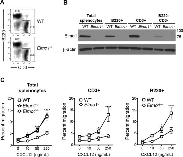
Figure 1. Impaired migration of Elmo1−/−splenocytes.
(A) Splenocytes from WT and Elmo1−/− mice were labeled with anti-B220 and anti-CD3 and analyzed by flow cytometry. The percentage of B220+ and CD3+ cells among all live splenocytes (7-AAD−) is shown. (B) Splenocytes were labeled as in A, FACS-sorted and analyzed by IB with antibodies indicated (left). Relative molecular weights are shown (right). One representative experiment of five is shown for A and B. (C) Transwell migration of splenocytes from WT, Elmo1+/− and Elmo1−/− mice to indicated concentrations of CXCL12. The percent of CD3+ and B220+ splenocytes that migrated to the lower chamber was determined by flow cytometry (n = 6 mice/genotype, ±SEM).

