
Figure 1.
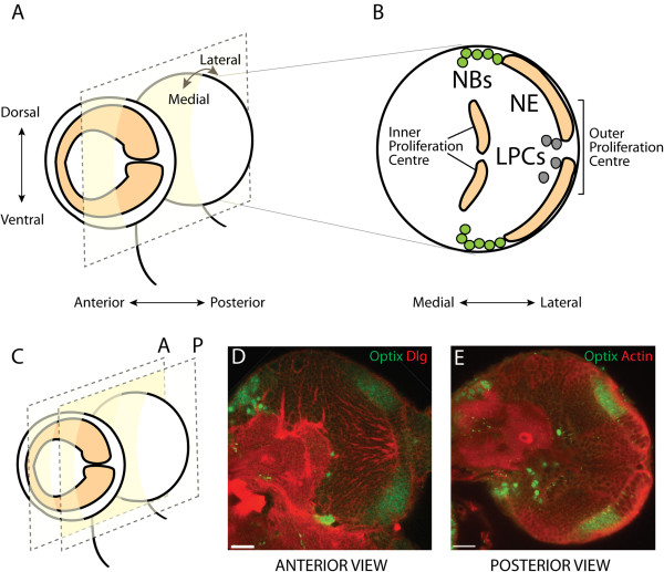
Optix is expressed in half of the optic lobe neuroepithelium. (A) Cartoon of a lateral view of larval third instar brain lobes. The optic lobe outer proliferation centre (OPC) is a horseshoe-shaped neuroepithelium (orange), which covers the lateral side of each brain lobe. A frontal cross-section is indicated by the dotted square. Dorsal-ventral, anterior-posterior and medial-lateral axes indicated by arrows. (B) Cartoon of a posterior frontal cross-section through a brain lobe at mid third instar. The OPC neuroepithelium (NE) generates two kinds of neural precursor: asymmetrically dividing medulla neuroblasts (NBs, green) and lamina precursor cells (LPCs, grey). Incoming retinal axons in the optic nerve enter through the central gap in the neuroepithelium. Medial-lateral axis indicated by arrows. (C) Cartoon of a lateral view from (A) showing the planes of two frontal cross-sections (dotted squares), one anterior (A) and one posterior (P). (D) Anterior confocal cross-section through a brain lobe at mid third instar. Cells are outlined in red by Discs large (Dlg) staining. Optix protein (green) is expressed across the neuroepithelium with a central gap. (E) Posterior confocal cross-section through a brain lobe at mid third instar. Cells are outlined in red by Actin staining (Phalloidin). Optix protein (green) is symmetrically expressed across the neuroepithelium with a sharp boundary.