
Figure 7.
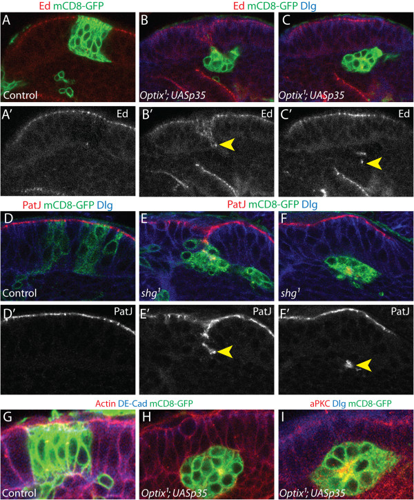
Loss of adhesion leads to ectopic neuroepithelial rosette formation. (A-C’) Cell death in Optix1 null mutant MARCM clones was rescued by expression of p35, which inhibits apoptosis. Clones are labelled with mCD8-GFP (green), Echinoid labels adherens junctions (Ed, red), cells are outlined by Dlg (blue). (A-A’) Control clones expressing p35 remain in the neuroepithelium. (B-C’)Optix1 mutant clones expressing p35 form ectopic neuroepithelial rosettes in the underlying medulla cortex. (D-F) Null mutant clones for DE-Cadherin (shg1) delaminate basally from the neuroepithelium to form rosettes below in the underlying medulla cortex (E, F), in contrast to FRTG13 control clones. (D) Clones are labelled with mCD8-GFP (green), PatJ labels adherens junctions (PatJ, red), cells outlined by Dlg (blue). (G-I) Apically localised proteins cluster at the centre of the neuroepithelial rosettes formed by rescued Optix1; UASp35 clones. (G-H) Clones are labelled with mCD8-GFP (green), DE-Cadherin labels adherens junctions (DE-Cad, blue), cells outlined by Phalloidin (F-Actin, red). (I) Clone is labelled with mCD8-GFP (green), cells outlined by Dlg (blue), aPKC is an apically localised protein (aPKC, red). (A-I) Posterior cross-sections are shown in all images. The apico-basal axis of the neuroepithelium is oriented vertically in each image, with the apical surface at the top and the basal surface at the bottom.