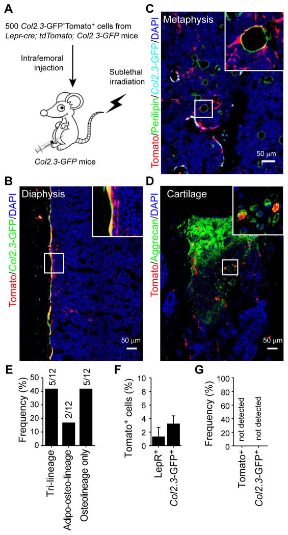
Figure 6. LepR+ cells give rise to osteoblasts, adipocytes, and chondrocytes after intrafemoral transplantation.
(A) Experimental design.
(B–D) Representative femur sections from Col2.3-GFP mice transplanted with 500 Tomato+Col2.3-GFP− cells as described in (A) (n=5). Note that the transplanted Tomato+Col2.3-GFP− cells gave rise to Col2.3-GFP+ osteoblasts (B), Perilipin+ adipocytes (C), and Aggrecan+ cartilage cells (at the injection site, D).
(E) Fraction of recipient mice in which Tomato+ cells were observed to contribute to each of the indicated mesenchymal lineages (n=12 mice).
(F) The percentage of LepR+ bone marrow stromal cells or Col2.3-GFP+ osteoblasts that were also Tomato+ (donor-derived) in the femurs of recipient mice (mean±SD from 3 mice in 3 independent experiments).
(G) No Tomato+LepR+ cells or Tomato+Col2.3-GFP+ cells were observed in the femurs of mice transplanted with 105 non-hematopoietic Col2.3-GFP−Tomato− cells from Lepr-cre; tdTomato; Col2.3-GFP mice (n=3).

