Fig. 1.
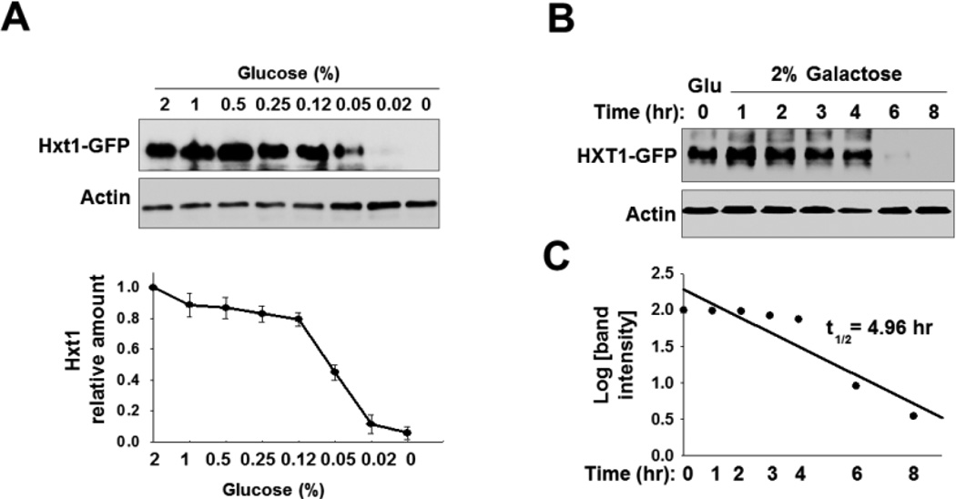
Hxt1 protein levels are posttranslationally downregulated in response to glucose starvation. (A) Western blotting analysis of the expression levels of Hxt1-GFP in the plasma membrane-enriched fraction. Yeast cells (WT) expressing Hxt1-GFP were grown in SC-2% glucose medium to mid log phase (O.D600nm = 1.2–1.5) and equal amounts of cells were shifted to SC medium containing different glucose concentrations. Membrane fractions were immunoblotted with anti-GFP antibody (top panel), and the intensity of each band on the blot was quantified by densitometric scanning (bottom panel). (B) Yeast cells (WT) expressing Hxt1-GFP were grown in SC-2% glucose (Glu) medium to mid log phase and equal amounts of cells were shifted to SC-2% galactose medium and incubated for indicated times. Membrane-enriched fractions were immunoblotted with anti-GFP antibody. (C) Determination of the half-life of Hxt1-GFP protein. The Western blot images in (B) were scanned and the half-life was determined, as described in the materials and methods.

