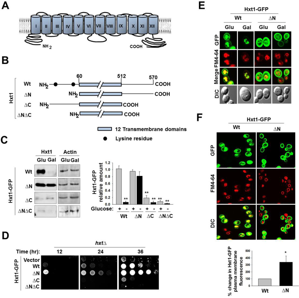
Fig. 3.
The N-terminal cytoplasmic domain of Hxt1 is required for its turnover. (A) Schematic diagram of predicted secondary structure of Hxt1. Twelve transmembrane domains and cytosolic N- and C-terminal tails are shown. (B) Schematic maps of Hxt1 constructs (Wt, 60–570 aa (ΔN); 1–512 aa (ΔC); 60–512 aa (ΔNΔC)) showing lysine residues at its N-terminal domain. (C) Yeast cells (WT) expressing indicated Hxt1-GFP proteins were grown as described in Fig. 2A, and plasma membrane fractions were immunoblotted with anti-GFP antibody (left panel), and the intensity of each band on the blot was quantified by densitometric scanning (right panel, **P < 0.001). Actin was served as loading control. (D) Yeast cells (hxtΔ) expressing indicated Hxt1-GFP proteins were spotted on 2% glucose plate supplemented with Antimycin A (1µg/ml). The first spot of each row represents a count of 5 × 10⁷ cell/ml, which is diluted 1:10 for each spot thereafter. The plate was incubated for indicated times and photographed. (E) Yeast cells (WT) expressing indicated Hxt1-GFP proteins were grown as described in Fig. 2A and analyzed by confocal microscopy. (F) Yeast cells (WT) expressing indicated Hxt1-GFP proteins were grown in SC-2% glucose medium to mid log phase and stained with FM6-64 (red). Confocal microscope images (top panel) and quantification of relative fluorescent intensity of Hxt1-GFP at the plasma membrane (bottom panel, * P <0.05) were shown.

