Fig. 5.
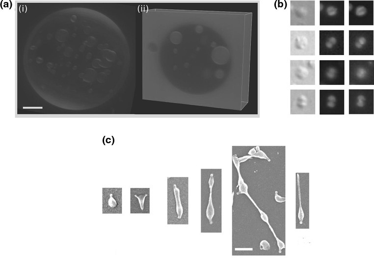
Binary fission of cells driven by ESCRT/Cdv or mechanical forces. a Three-dimensional reconstruction of ESCRT-III treated giant unilaminar vesicles (GUV). i Membrane staining of the intra-vesicular bodies that were formed after reconstitution of the eukaryote ESCRT-III complex on the outside of a GUV. ii Z-stack confocal image of the same GUV showing that the intra-vesicular bodies were filled with the extra-vesicular content as a result of ESCRT-III-induced inward budding. Scale bar 5  . b In situ immunofluorescence localization of CdvA (red middle column) and CdvB (green right column) during the constriction of four S. acidocaldarius cells. Note that both CdvA and CdvB are localized between the two segregated chromosomes (blue stained with DAPI). Left column phase-contrast illumination. c Stages in the division of
  M. genitalium. Newborn cells possess a single terminal organelle. Division starts when the terminal organelle duplicates, leading to second organelle at the other cell pole. Cytoskeleton filaments then elongate the cell, and create a constricted tube at the cell middle. After the formation of a chain of filaments, some cells are torn off the chain and can start a new reproduction cycle. Scale bar 500 nm. a Modified with permission from reference Wollert et al. (2009)
  (2009) Nature Publishing Group. b Modified with permission from reference Samson et al. (2008)
  (2008) National Academy of Sciences, USA. c Modified with permission from reference Lluch-Senar et al. (2010)
  (2010) John Wiley and Sons, Inc. (Color figure online)

