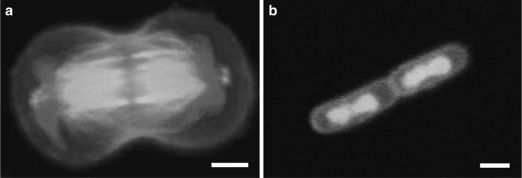
Fig. 1.

Segregation of the duplicated genomes to both cell halves during cell division. a Chinese hamster ovary cell in anaphase. The genome is labeled in blue while actin and microtubules are seen in red and green, respectively. Scale bar 5 μm. Image courtesy of Ahna Skop, UW-Madison. b E. coli bacterium that is just about to finish cell division. The genome is stained in green; the cell envelope in red. Scale bar 1 μm. Image courtesy of Fabai Wu, TU Delft. (Color figure online)
