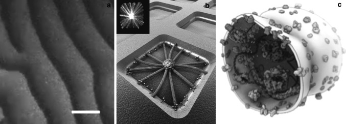
Fig. 4.

Examples that illustrate how in vitro synthetic biology can help to unravel the mechanism of the cell-division machinery. a Waves of Min proteins (involved in bacterial cell division of E. coli) on a supported lipid bilayer, from Loose et al. (2008). Orange and green color denotes MinE and MinD proteins, respectively. Scale bar is 50 μm. b Artist’s impression of microtubule asters (involved in eukaryotic cell division) that are positioned by cortical motor proteins in microfabricated chambers. Chamber size is 15 μm. Image courtesy Marileen Dogterom. The top left inset shows the experimental result, from Laan et al. (2012). c Artistic sketch that illustrates the future dream of a vesicle with reconstituted components with an engineered minimal divisome that will cause the vesicle to divide. Image courtesy Bert Poolman. (Color figure online)
