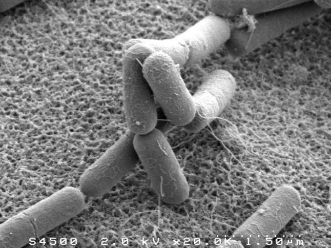
Fig. 1.

Electron microscopy images of B. subtilis, taken by Thierry Meylheuc. In a given growth medium, the different bacterial cells have a smooth, highly reproducible cylindrical shape, with relatively small fluctuations in length and radius. The image is reproduced from Chastanet and Carballido-Lopez (2012), courtesy of A. Chastanet
