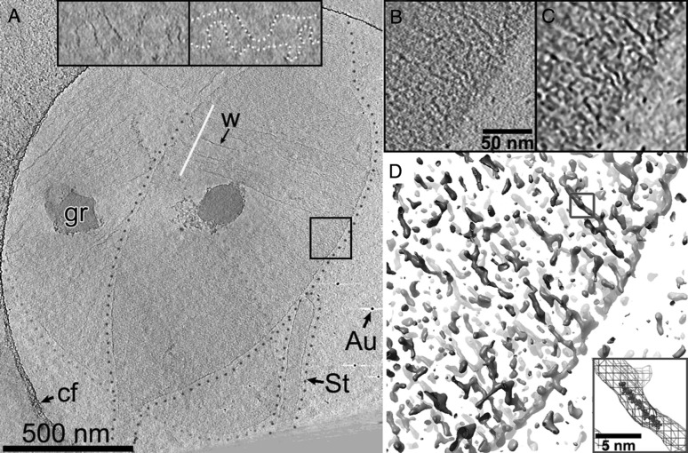
Fig. 2.
Details of peptidoglycan organization as obtained by cryo-electron tomography; reproduced from Gan et al. (2008), courtesy of Grant J. Jensen. Computational reconstructions of the three-dimensional electron density of a cell-wall sacculus of the Gram-negative bacterium C. crescentus reveal a circumferential orientation of the cell-wall glycan strands: shown are two overlapping cell-wall sacculi (a), outlined by green or violet dotted lines. The boxed region in a is magnified in b–d. d An iso-density plot, which shows long circumferentially oriented structures that are presumably individual glycan strands. The inset in d displays a superposition of the blue-boxed glycan strand and an atomic model of a nine-subunit-long glycan strand, for comparison of scale. (Color figure online)

