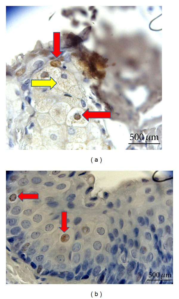
Figure 6.

BrdU labeled MSCs were shown to infiltrate the meibomian glands and conjunctival epithelium. (a) Meibomian gland infiltration of BrdU labeled MSCs. Cells with BrdU+ stained nucleus (red arrows). Spread of BrdU stained areas to the cytoplasm of some cells (yellow arrow). (b) BrdU+ cells in the conjunctival epithelium (red arrow) (Leica HMLB45: Germany, 2000; magnification: ×400).
