Figure 1.

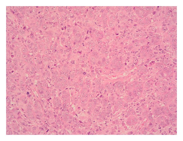
Hematoxylin and eosin stained section of cerebellar lesion at 20x magnification showing general cytoarchitectural features of the tumour.

Hematoxylin and eosin stained section of cerebellar lesion at 20x magnification showing general cytoarchitectural features of the tumour.