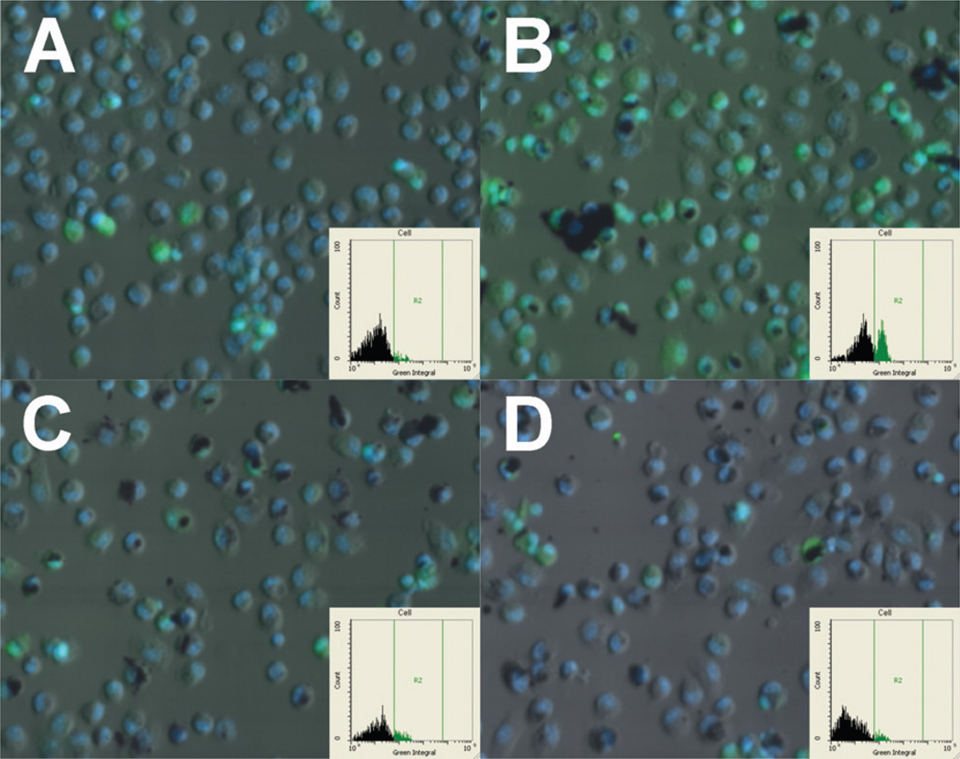
Figure 9.
Computer generated (iCys) images of C57BL/6 alveolar macrophages exposed to FAM FLICA green Caspase-1 substrate. (A) No-particle control cells at 3 h. (B) Cells exposed to raw MWCNT for 3 h. (C) Cells exposed to functionalized raw MWCNT for 3 h. (D) Cells exposed to functionalized pure MWCNT for 3 h. Green fluorescence indicates Caspase-1 signal. Blue fluorescence is the nucleus. Insets show gated positive signal for Caspase-1 (only present in panel B).

