
Figure 4. Subcellular localization of GFP-AR(294–556) and GFP-NTD in LAPC4, C4-2 and LNCaP cells.
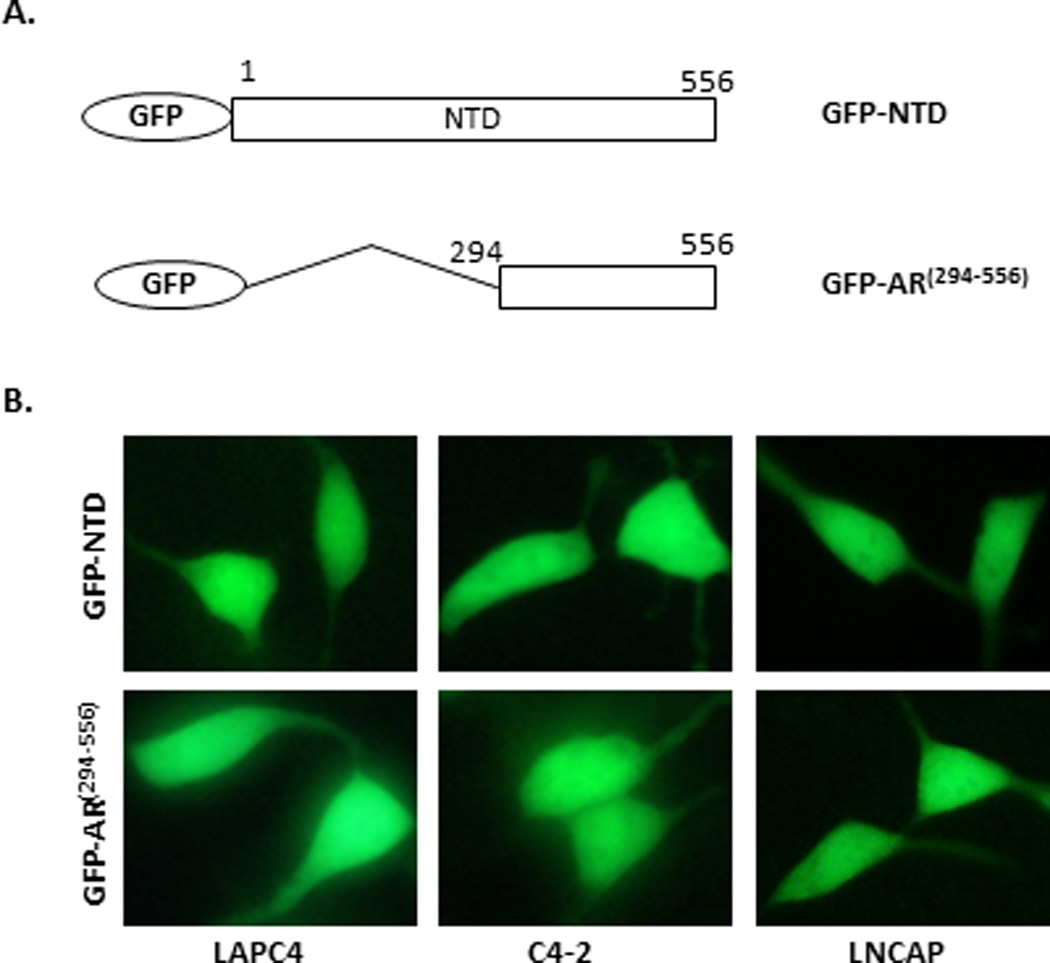
(A) Diagram of GFP fusion constructs GFP-NTD and GFP-AR(294–556). (B) Representative images of GFP-NTD and GFP-AR(294–556) in transiently transfected LAPC4, C4-2, and LNCaP cells. The subcellular localization was assessed in complete medium by fluorescence microscopy after 16 hours of transfection. The experiment was reproduced 5 times.