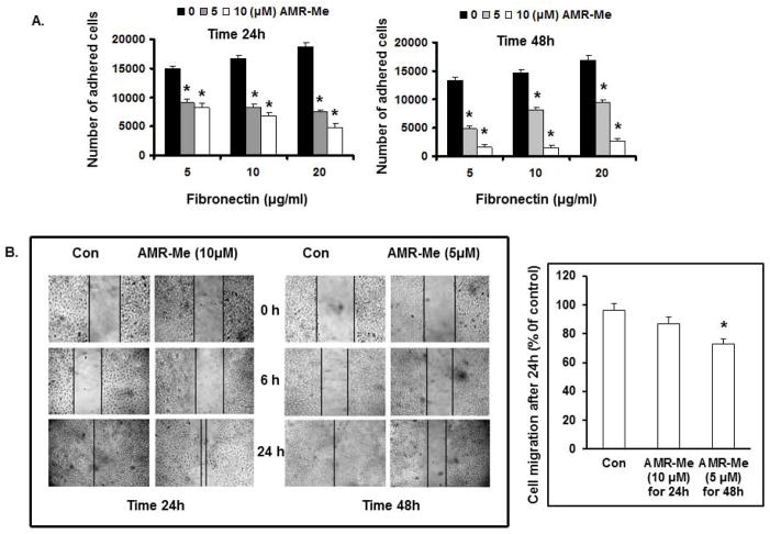
Fig. 2.
Effect of AMR-Me on adhesion and migration of B16F10 cells. (A) Inhibition of FN-induced cell adhesion by AMR-Me (10 μM) for 24 h and AMR-Me (5 μM) for 48 h. Inhibition of cell adhesion by AMR-Me was significant (P<0.001) compared to vehicle control with increasing doses of FN. (B) Inhibition of cell migration by treatment with AMR-Me (10 μM) for 24 h and AMR-Me (5 μM) for 48 h compared to vehicle control as evidenced at different time intervals (0, 6 and 24 h). After 24 h incubation migrated cells across the black lines were counted in six random fields from each treatment and the mean number of cells in the marked zone was quantified by three independent experiments. The graphical representation in terms of % of control exhibited AMR-Me (5 μM) for 48 h compared to vehicle control caused significant inhibition (*P<0.05) of cell migration.

