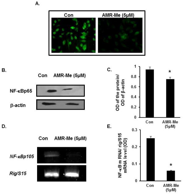
Fig. 7.
Inhibition of nuclear translocation and expression of NF-κB following AMR-Me treatment. (A) Immunocytochemistry showing inhibition of nuclear localization of NFkBp65 with AMR-Me (5 μM) for 48 h with respect to vehicle control. (B) Western blot showing reduced expression of NF-κBp65 in nuclear fraction of B16F10 melanoma cells treated with AMR-Me (5 μM) for 48 h with respect to vehicle control. (C) The respective band intensities of NF-κBp65 as calculated by Image J software exhibiting significant inhibition (*P<0.05) of pNF-κBp65 in nuclear fraction of B16F10 melanoma cells treated with AMR-Me (5 μM) for 48 h with respect to vehicle control. (D) RT-PCR showing downregulated expression of NF-κBp105 in B16F10 melanoma cells treated with AMR-Me (5 μM) for 48 h with respect to vehicle control. (E) The respective band intensities as calculated by Image J software exhibiting significant inhibition (*P<0.01) of NF-κBp105 in melanoma cells treated with AMR-Me (5 μM) for 48 h with respect to vehicle control.

