
Figure 7.
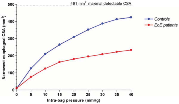
Functional luminal imaging in eosinophilic esophagitis quantified remodeling effects of the esophagus. Esophageal distensibility plots in control subjects (blue) and eosinophilic esophagitis (red) demonstrating diminished distensibility for distension pressures above 5 mm Hg. The calculated value for constant cross sectional area in spite of increasing distension pressure is used to generate the distensibility plateau (DP). Data from Kwiatek, M.A., et al., Mechanical properties of the esophagus in eosinophilic esophagitis. Gastroenterology, 2011. 140(1): p. 82–90.