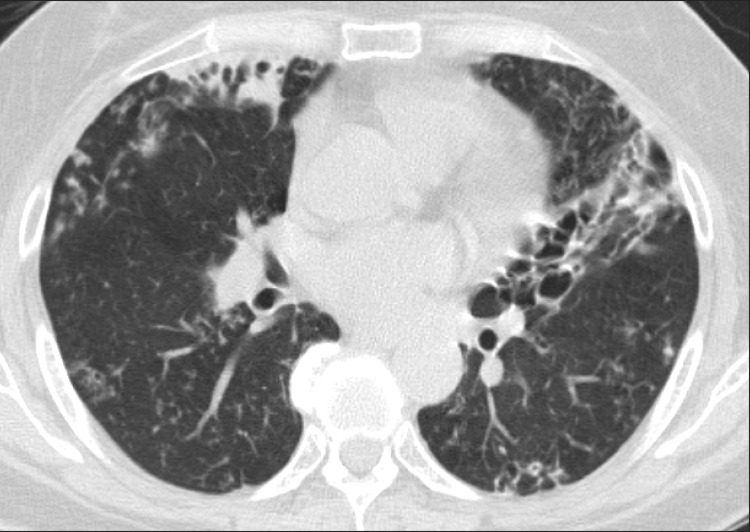
Figure 2.
Nodular bronchiectatic form of nontuberculous mycobacterial lung disease in a 60-year-old female with Mycobacterium abscessus lung disease. Chest high-resolution computed tomography shows severe bronchiectasis in the right middle lobe and the lingular segment of the left upper lobe. Note the multiple small nodules suggesting bronchiolitis in both lungs.

