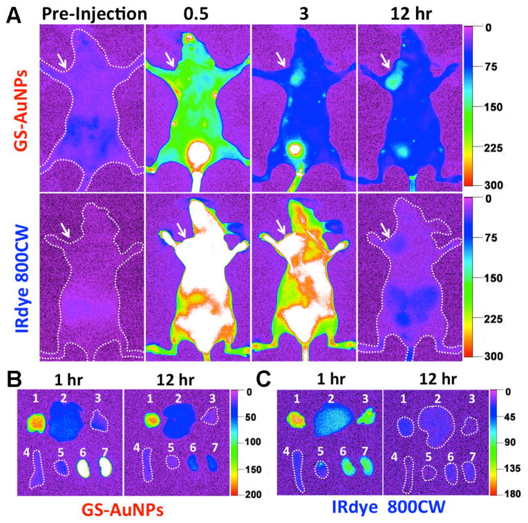
Figure 2.
In vivo and ex vivo NIR fluorescence imaging of MCF-7 tumor-bearing mice IV injected with GS-AuNPs and IRdye 800CW. A) Representative in vivo NIR fluorescence images collected at p.i. time points of 0.5, 3, and 12 hr, respectively. The tumor areas were indicated with arrows. B, C) Ex vivo fluorescence images of organs and tumors taken from the MCF-7 tumor-bearing mice IV injected with GS-AuNPs (B) and IRdye 800CW (C) at the time points of 1 and 12 hr p.i., respectively. In each image: 1, tumor; 2, liver; 3, lung; 4, spleen; 5, heat; 6, kidney (left); 7, kidney (right). More images related to the tumor targeting of the IRdye 800CW were shown in Figure S5.

