
Figure 6. Perivascular glioma cells do not compromise vascular smooth muscle cell function.
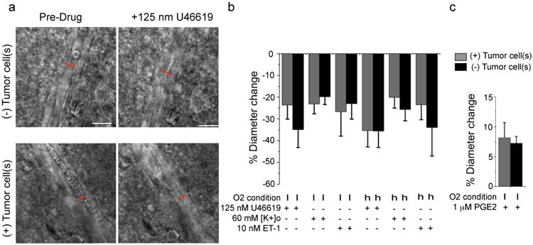
DIC images of vessels without (top) or with (bottom) perivascular glioma cells before and after application of 125 nM U46619. Perivascular glioma cell presence was verified by eGFP-fluorescence (a). Average change in vessel diameters observed at high (h) (95%) and low (l) (20%) oxygen for vessels associated (grey) and not associated (black) with glioma cells when exposed to 125 nM U46619, 60 mM [K+]o, 10 nM ET-1 (b) and 1 μM PGE2 (c). For 1 μM PGE2, arterioles were preconstricted with 125 nM U46619 for 20 min. Statistical data as follows: 20% Oxygen. 125 nM U46619: (+)Tumor Cell(s) n=11, (-)Tumor Cell(s) n=11, two-tailed Mann Whitney Test, p=0.40; 60 mM [K+]o: (+)Tumor Cell(s) n=10, (-)Tumor Cell(s) n=9, two-tailed unpaired t-test, p=0.57; 10 nM ET-1: (+)Tumor Cell(s) n=8, (-)Tumor Cell(s) n=9, two-tailed unpaired t-test, p=0.78; 1 μM PGE2: (+)Tumor Cell(s) n=14, (-)Tumor Cell(s) n=21, two-tailed unpaired t-test, p=0.72. 95% Oxygen. 125 nM U46619: (+)Tumor Cell(s) n=14. (-)Tumor Cell(s) n=12, two-tailed unpaired t-test, p=0.99; 60 mM [K+]o: (+)Tumor Cell(s) n=11, (-)Tumor Cell(s) n=19, two-tailed Mann-Whitney Test, p>0.9999; 10 nM ET-1: (+)Tumor Cell(s) n=7, (-)Tumor Cell(s) n=9, two-tailed unpaired t-test, p=0.48). Error bars refer to SEM Scale, 20 μm.