
Figure 8. Perivascular glioma cells disrupt astrocyte-mediated vascular coupling.
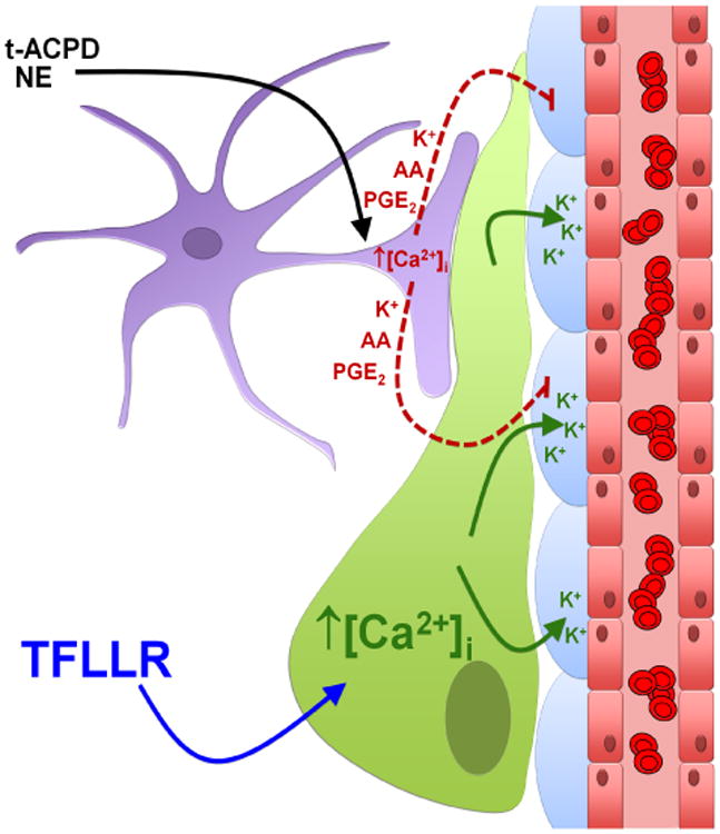
The perivascular glioma cell (green) inserts itself between the vascular smooth muscle cells (VSMCs) (blue) surrounding the vascular endothelial cells (red), displacing the astrocytic endfoot (purple). Bath application of norepinephrine (NE) and trans-ACPD (t-ACPD) activates astrocytic G-protein coupled receptors causing an increase in astrocytic [Ca2+]i. Due to perivascular glioma cell displacement of the astrocytic endfoot, astrocyte-released vasoactive molecules (arachidonic acid (AA), prostaglandin E2 (PGE2), and K+) can no longer reach the VSMCs to cause alterations in vessel diameter. Glioma cell-protease activated receptor 1 (PAR1) activation by the artificial ligand, TFLLR, causes increases in glioma cell [Ca2+]i, which activates Ca2+-activated K+ channels. Glioma efflux of K+ onto VSMCs causes vasoconstriction of arterioles.